Owner
A C
A C
A common observation of mast cell tumours is that they often fluctuate in size.
A mast cell is a type of blood cell that is normally involved in allergic or inflammatory responses. A mast cell tumor is one of the most common skin cancers in dogs but can also be found in the liver, spleen, or bone marrow.
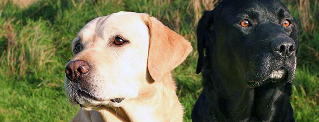
Breeds commonly affected by mast cell tumors:
Mast cells are more commonly seen in older dogs, however, can occur in younger dogs as well.
Mast cell tumors can vary in appearance.
Common signs of a mast cell tumor:
Common mast cell tumor behaviors:
They are typically locally invasive but can spread as the grade of tumor increases.
Diagnosis is based on a fine needle aspirate or biopsy of the lump tissue. Other tests such as bloodwork, urine analysis, bone marrow biopsy, or ultrasound may be required to determine the extent of the disease and whether it has spread to other parts of the body such as the local lymph nodes, spleen, or liver.
Common diagnostic tests:
| Grade 1 | localized and rarely spread to other parts of the body |
| Grade 2 | extend to underlying tissue and approximately 20% chance of spreading |
| Grade 3 | difficult to isolate a tumor from underlying tissues and approximately 80-100% chance of spreading |
Depending on the results of the biopsy, treatment may involve:
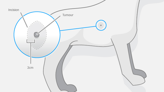
Most cases of mast cell tumors are resolved with surgery. Mast cell tumors are locally invasive, meaning they spread into the surrounding tissue. For this reason, a large surgical incision around and below the tumor is removed to help capture all the cancer cells. Unfortunately, it cannot be guaranteed that all the cancer cells are removed due to the deceptive nature of the cancer cells. If regrowth occurs, further surgery, radiation, chemotherapeutic, or toceranib treatment is recommended.
Tumors that are considered Grade 1 to 2 will have a low incidence of spreading whereas a Grade 3 tumor is more likely to spread. In these more serious cases where a tumor may be considered too big or too difficult to surgically remove or it has spread already, chemotherapy, toceranib, additional surgery and/or radiation therapy may be recommended.
The prognosis is dependent on tumor grade, stage, and the ability to fully remove a tumor surgically. Grade 1 and 2 tumors are likely to be cured if removed adequately by surgery. In cases where margins are incomplete, there is a 20-30% chance of recurrence.
Unfortunately, affected dogs are at a higher risk for developing further primary mast cell tumors and early detection is extremely important for the success of treatment.
Dogs that are diagnosed with mast cell tumors that have already spread to other parts of the body have a poor prognosis. In these cases, treatment is aimed at maintaining a good quality of life.
London CA, et al. Expression of stem cell factor receptor (c-kit) by the malignant mast cells from spontaneous canine mast cell tumours. J Comp Path 1996;115:399-414.
Bookbinder PF, Butt MT, Harvey HJ. Determination of the number of mast cells in lymph node, bone marrow, and buffy coat cytologic specimens from dogs. J Am Vet Med Assoc 1992;200:1648-50.
Gieger TL, et al. Biologic behavior and prognostic factors for mast cell tumors of the canine muzzle: 24 cases (1990-2001). J Vet Intern Med 2003;17:687-92.
Murphy S, et al. Relationships between the histological grade of cutaneous mast cell tumours in dogs, their survival and the efficacy of surgical resection. Vet Rec 2004;154:743-6.
Séguin B, Leibman NF, Bregazzi VS, et al. Clinical outcome of dogs with grade-II mast cell tumors treated with surgery alone: 55 cases (1996-1999). J Am Vet Med Assoc 2001;218:1120-3.
Cahalane AK, Payne S, Barber LG, et al. Prognostic factors for survival of dogs with inguinal and perineal mast cell tumors treated with surgery with or without adjunctive treatment. 68 cases. J Am Vet Med Assoc 2004;225:401-8.
Simpson AM, Ludwig LL, Newman SJ, et al. Evaluation of surgical margins required for complete excision of cutaneous mast cell tumors in dogs. J Am Vet Med Assoc 2004;224:236-40.
Frimberger AE, Moore AS, LaRue SM, et al. Radiotherapy of incompletely resected, moderately differentiated mast cell tumors in the dog: 37 cases (1989-1993). J Am Anim Hosp Assoc 1997;33:320-4.
Hahn KA, King GK, Carreras JK. Efficacy of radiation therapy for incompletely resected grade-III mast cell tumors in dogs: 31 cases (1987-1998). J Am Vet Med Assoc 2004;224:79-82.
Thamm DH, Mauldin EA, Vail DM. Prednisone and vinblastine chemotherapy for canine mast cell tumor--41 cases (1992-1997). J Vet Intern Med 1999;13:491-7.
Rassnick KM, Moore AS, Williams LE, et al. Treatment of canine mast cell tumors with CCNU (lomustine). J Vet Intern Med 1999;13:601-5