
2024 Ultimate Guide to Dog Anatomy
As the pace of veterinary advancement accelerates, even the most experienced veterinary teams are challenged to keep up with all the changes that impact their practice. Veterinary teams need practical, concise and relevant visual aids at their fingertips while in practice, helping them to prescribe the right information at the right time, to improve client communication, increase compliance rates, enhance the pet owner experience and most importantly better pet health outcomes.
Why visual aids are important.
 Your working day is often fast-paced and always changing. But, a large part of your job is pet owner communication.
There are many barriers to managing client communication in a veterinary setting:
- Veterinary support teams find it difficult to juggle work and keep up to date with the latest pet health information
- Pet owners struggle to describe the pet's symptoms
- Pet owners are often pre-occupied with pet restraint or their children
- Veterinarians may not always have the time they need to fully understand a case
- The primary carer is not always present
- There is never enough time to explain everything from diagnosis, treatment plan, prognoses and more
A simple way to overcome these barriers is to use digital visual aids and digital pet treatment summaries.
VetCheck is going to help you be the best vet or nurse clinician you can be by:
- Offering you a proven client communication and engagement system that helps you deliver value and high quality care
- Giving you access to up to date pet health information e.g. health updates, treatment options, potential outcomes and prognoses and tools e.g. anatomical diagrams, clinical formulas and programs and home care videos
- Enhancing the delivery of your healthcare messages, managing client expectations and improving compliance for better pet health outcomes
- Improving your clinical efficiency and streamlining the care you deliver by improving patient data capture and reducing call backs
Needing veterinary visual aids, anatomical diagrams, home compliance videos and images to improve the quality of care that you can provide?
Anatomical terminology
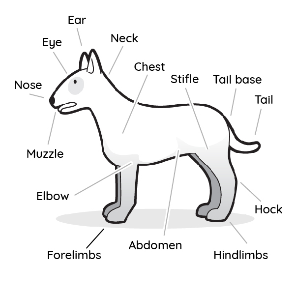
The use of veterinary anatomical terminology can be confusing. When discussing a pet's condition, always use both technical and laymens terminology. People think and hear in pictures. Below are a selection of visual aids to help you communicate the importance of the pet's health as well as the recommended veterinary services.
Get access to 100's of anatomical diagrams. Simply register online and once approved, gain access within a few minutes.
JOIN NOW
Common anatomical terminology
Here are some common veterinary terms and their meanings:
| Abdomen | Tummy |
| Dew claw | First digit |
| Patella | Knee cap |
| Stifle | Knee |
| Thorax | Chest |
| Digit | Finger or toe |
| Flank | Side of the body between chest and tail base |
| Muzzle | Nose and upper and lower lip |
| Pinna | Ear flap |
| Tarsus | Hock |
Pet senses
Pets communicate in a very different way than people do. They have the same basic senses like sight, hearing, smell, touch, and taste, but they use them differently to communicate with the world. In general, pets have a much better sense of smell, hearing, and sight than humans. This allows them to identify odours better, to hear noises at greater distances, and to see in the dark. Pets also have sharp teeth and claws that developed to help them survive in the wild.
In the wild, dogs are pack animals that require a strong leader. Their excellent senses of smell and hearing have allowed them to survive and catch prey in the wild. Because of their highly developed senses they are great trackers. Dogs identify each other by their unique scent. They have scent glands located around their bottom and use them to mark territory as their own. This is why we commonly see dogs greeting other dogs by sniffing their bottom.
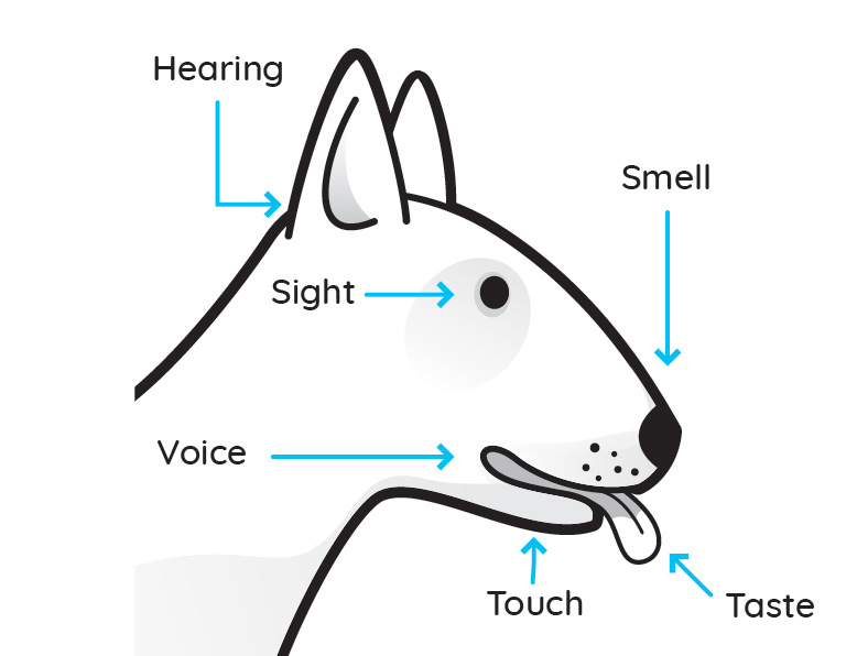
| Hearing | Dogs have a greater hearing range than people do. They can detect sound as low as 16 Hz frequency to as high as 100,000 Hz (people hear 20 to 20,000 Hz ). Their ears have a great degree of flexibility that allows them to funnel sounds and easily locate the direction of sound. They can hear sounds much sooner and at much greater distances than people do. Dogs with cone ears naturally hear better than those with floppy ears. |
| Sight | People used to think that a dog’s sight is dichromatic (see in black and white). But the latest research suggests that dogs may actually see some color, though certainly not as much as people do. Depending on the dog breed, their field of vision can vary up to 270 degrees for sight hounds like greyhounds and whippets, and as low as 180 degrees for flat-faced breeds like the bulldog or Boston terrier. People also have a narrow field of vision of 180 degrees. Dogs can see much better at night than people do. Their eyes are more sensitive to light and motion than ours. They have a structure, the tapetum lucidum, that allows them to see in dim light. Have you ever noticed their eyes reflecting back when bright lights like a car headlight or flashlight are directed at them? |
| Voice | Different dog breeds have different voices. There are many different types of voices: bark, growl, howl, and whimper. A dog’s bark expresses different emotions like pleasure, fun, loneliness, fear, or stress. |
| Smell | Smell is the dogs’ primary sense. Dogs have nearly 220 million olfactory (smelling) cells, compared to 5 million in people. Dogs sniff to take in air quickly to identify different smells. Their sense of smell is extremely sensitive and the government often uses dogs to track people, drugs, or explosives. They can even use smell to sense human and animal moods such as fear, happiness, or sadness from long distances. |
| Taste | Dogs have 42 permanent teeth to chew on meat and vegetables. They have a broad tongue with only 1,700 taste buds, since their heightened sense of smell allows them to identify food. People have 9,000 taste buds. |
Get access to more anatomical diagrams on special senses.
- Normal eye
- Nusclear sclerosis
- Cataracts
- Glaucoma
- Corneal ulceration
- Normal ear
- Normal hearing aparatus
- Otitis externa
Simply register online and once approved, gain access within a few minutes.
JOIN NOWCardiovascular and Circulatory System
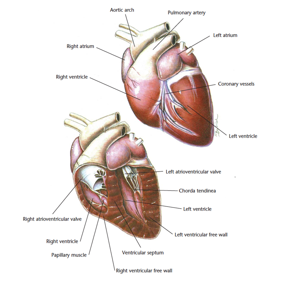
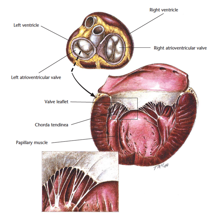
The cardiovascular system refers to the organs and vessels that allow blood to circulate nutrients, oxygen, carbon dioxide, wastes and hormones to the various cells within the body. The heart pumps oxygenated blood from the lungs to the rest of the body, while pumping deoxygenated blood to the lungs.
The heart is made up of the following structures:
- Aorta
- Pulmonary artery
- Right atrium
- Right ventricle
- Left atrium
- Left ventricle
- Ventricular septum
Cardiopulmonary Resuscitation (CPR) Guidelines
Download the VetCheck Cardiopulmonary Resuscitation (CPR) Guidelines for step by step instructions and videos.
REQUEST A FREE COPY NOWGood heart health starts with good nutrition.
Taurine, an important amino acid, is essential for strong heart muscles as well as eye and brain function.
Most commercial cat foods contain taurine.
But, in the case of homemade diets, cats are at higher risk of taurine deficiency and heart problems.
Cardiovascular conditions are particularly complex. In order for pet owners to make sound health decisions, they need to under the risks and benefits that come with medical treatments and diagnostic tests.
Studies have shown that people consider risk information easier to understand and recall when it is presented visually.
Get access to more anatomical diagrams on the cardiovascular system.
- Chronic Valvular Disease
- Heartworm Disease
- Feline Dilated Cardiomyopathy
- Feline Hypertrophic Cardiomyopathy
Simply register online and once approved, gain access within a few minutes.
JOIN NOWDigestive system
The digestive system is made up of the organs responsible for processing food into a format that can be used by the body in the form of energy and nutrients. Food enters the mouth and travels through the oesophagus, stomach, small intestine and large intestine before being passed through the anus as solid waste.
The digestive system includes the:
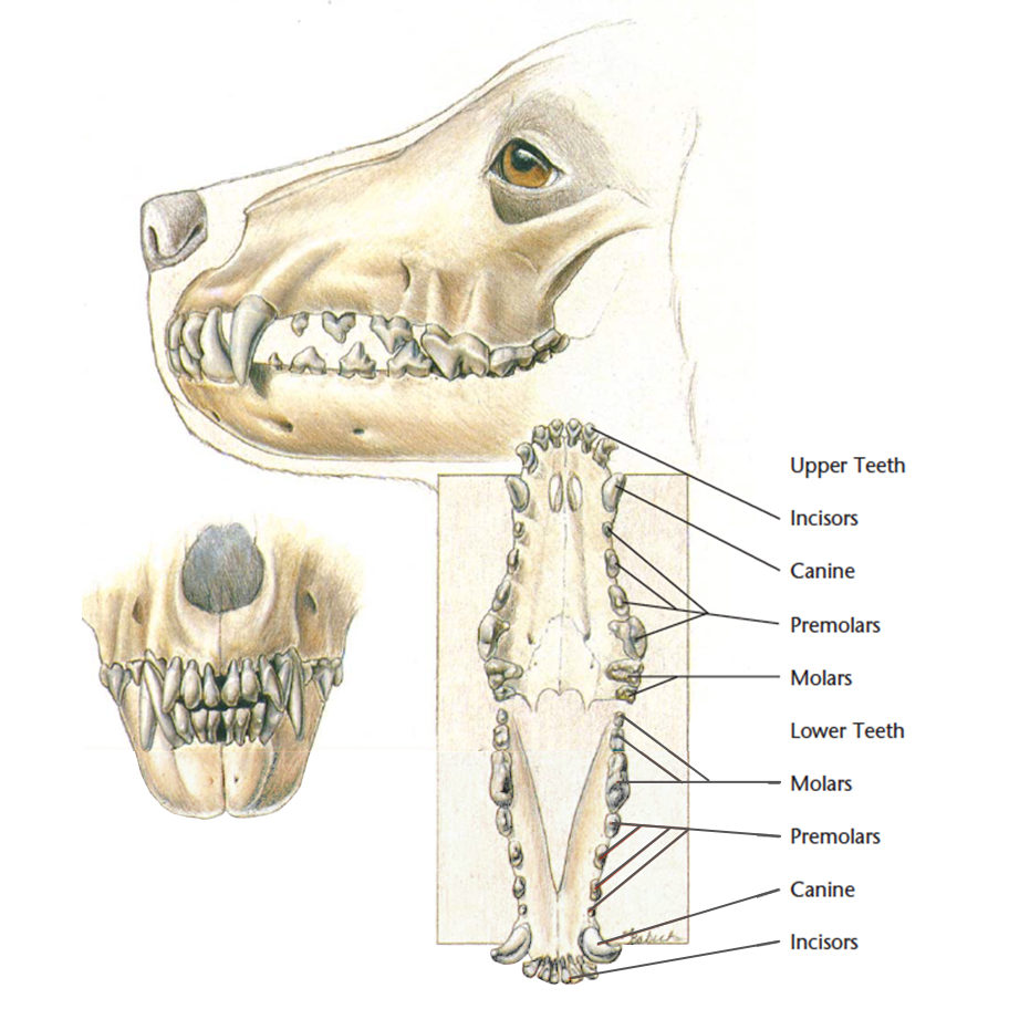
- Mouth & Teeth
- Tongue
- Salivary glands
- Oesophagus
- Stomach & Stomach Lining
- Small intestine
- Large intestine
- Pancreas
- Liver
- Gall bladder
Nutrition for good health
Dogs are omnivores meaning they need vegetables and meat. The ideal diet is one that is tailored to the individual nutritional requirements. This is based on the health, life stage and lifestyle.
An appropriate diet combines a high quality, balanced, commercial diet and human-grade foods. Unless you have specific recipes that have been formulated by a veterinary nutritionist, commercial diets should be considered, particularly for puppies up to 12 months of age. All reputable veterinary nutritional companies must follow strict dietary requirements to ensure the diets are balanced and nutritionally beneficial.
Mouth and teeth
Stomach and stomach lining
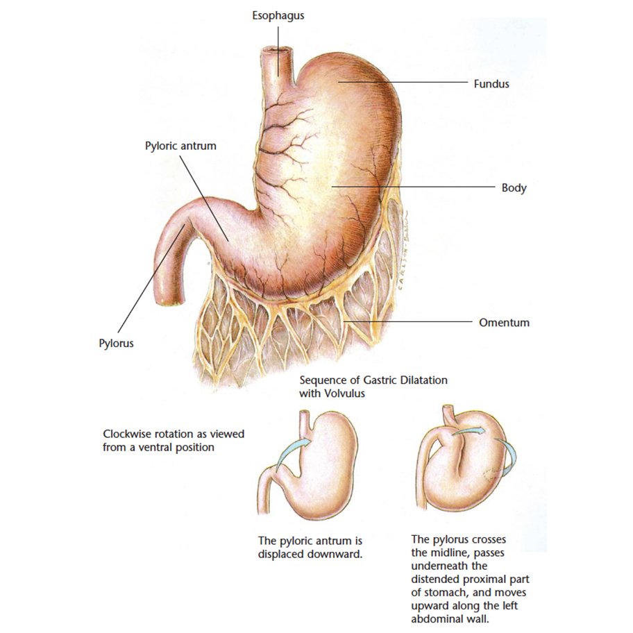
The food enters via the oesophagus, into the stomach is where the food is digested so that the nutrients can be absorbed.
Small intestine
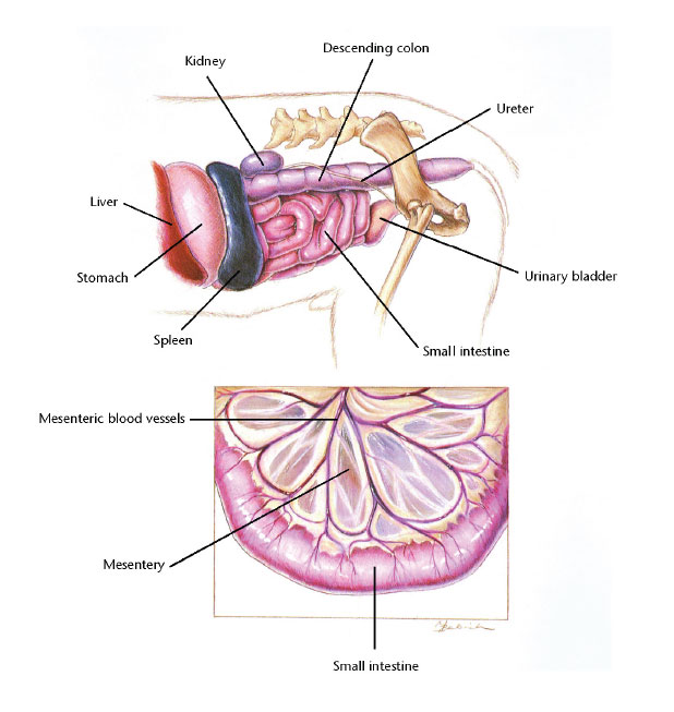
The small intestine connects the stomach to the large intestine. It can be broken into three sections - the duodenum, jejunum and ileum. It is where food absorption continues to take place after it has left the stomach.
Pancreas
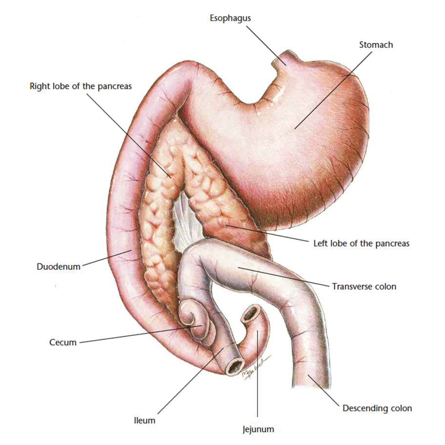
The pancreas is a gland located near the stomach. It produces a number of important hormones that aid in digestion and regulates blood sugar.
Liver
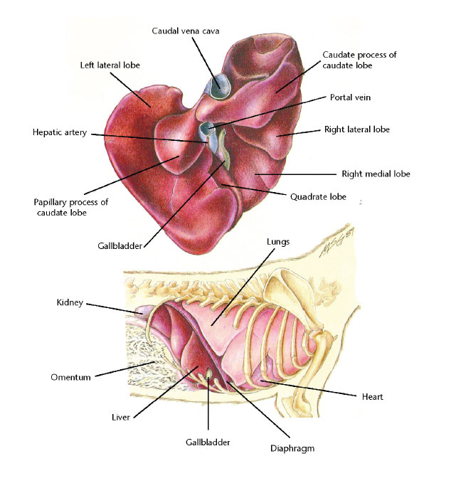
The liver is responsible for removing toxins that come from the digestive tract.
Get access to more anatomical diagrams on the digestive system.
- Hemorrhagic Gastritis with Ulcers
- Gastric Dilatation with Volvulus
- Intestinal Foreign Bodies
- Parvoviral Enteritis
- Intussusception
- Chronic Colitis
- Constipation/Colonic Impaction
- Acute Pancreatitis
- Exocrine Pancreatic Insufficiency
- End-Stage Liver Disease
- Hepatic Neoplasia
Simply register online and once approved, gain access within a few minutes.
JOIN NOWMusculoskeletal system
The musculoskeletal system is responsible for form, support, stability and movement. It is made up of skeletal bones, muscles, cartilage, tendons, ligaments, joints and connective tissue.
Common joints include the:
- Elbow
- Shoulder
- Hip
- Stifle (knee)
Shoulder
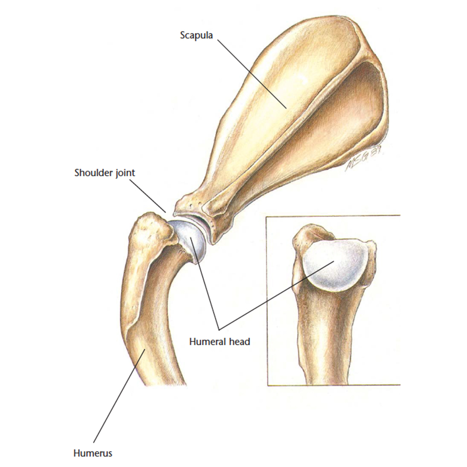
The shoulder joint is made up of the scapula (shoulder blade) and humerus (large arm bone).
VetCheck Passive Range of Motion Guidelines
Download the VetCheck Passive Range of Motion Guidelines for step by step instructions and videos.
DOWNLOAD NOWPelvis
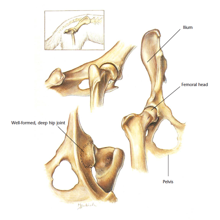
The pelvis is where the femoral (large leg bone) head fits into the hip joint.
Stifle and patella
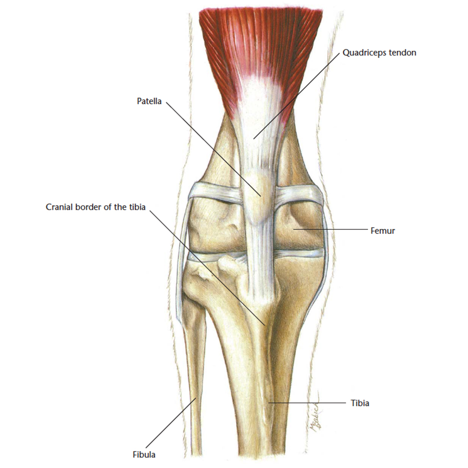
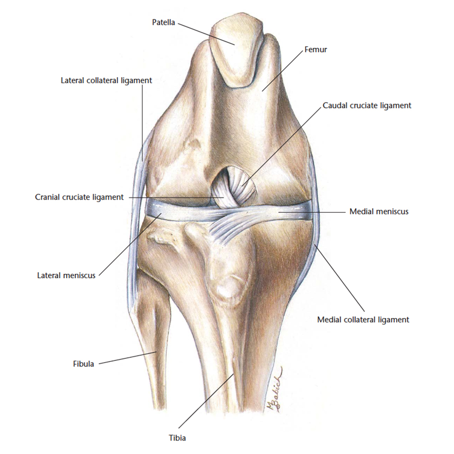
The stifle is the knee and the patella is the knee cap. They are both positioned in the hindlimbs of the cat.
Get access to more anatomical diagrams on the musculoskeletal system.
- Intervertebral Disk Disease
- Osteochondritis Dissecans
- Ununited Anconeal Process/Panosteitis
- Hip Dysplasia
- Femoral Fracture
- Ruptured Cranial Cruciate Ligament
- Patellar Luxation
Simply register online and once approved, gain access within a few minutes.
JOIN NOWRespiratory system
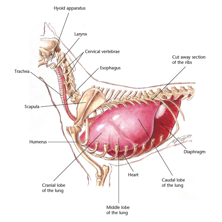
The respiratory system is responsible for bringing oxygen into the body and removing wastes in the form of carbon dioxide. Pets cannot regulate their heat through their skin in the form of sweat - the respiratory system is responsible for regulating the body temperature for example panting when the pet is hot.
The respiratory system includes the:
- Nose
- Pharynx
- Larynx
- Trachea
- Bronchi (smaller airways)
- Lungs
Get access to more anatomical diagrams on the respiratory system.
- Tonsillitis
- Collapsing Trachea
- Pulmonary Edema
JOIN NOW
Urogenital system
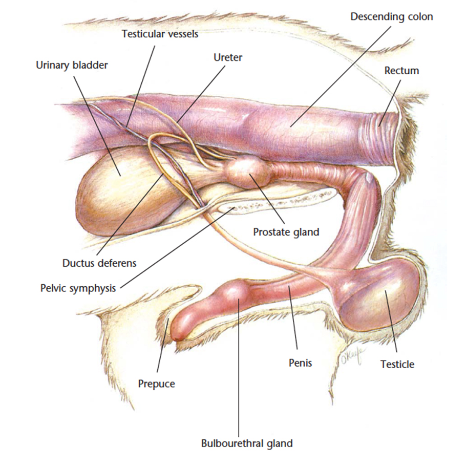
The urogenital system refers to the urinary system that includes the kidneys, ureter, urethra and bladder in the excretion of liquid wastes and the reproductive system that includes the female uterus, ovaries, fallopian tubes and vagina and the male testes, epididymis, vas deferens and penis.
Lower urinary tract
Female genitalia

A spey or ovariohysterectomy involves the removal of both ovaries and the uterus. Without desexing, your pet will proceed to puberty and leave bloodstains around the house during each heat cycle.
Benefits of desexing:
- Prevention of unwanted litters
- Health benefits for females such as womb infections (pyometra), breast cancer
- Health benefits for males such as reduced prostate disease, testicular cancer, perianal tumours
- Behavioural benefits such as reduced spraying, marking, fighting if castration occurs before 6 months of age or before the onset of these behaviours
- Prevention of hormonal changes that can interfere with the medical management of pets with diabetes or epilepsy
Desexing is usually recommended before puberty between the age of 4 and 9 months but can occur at any age. Six months is an ideal age as the puppy vaccination series is usually completed. Males undergo a castration which is the removal of both testicles from beneath the skin. Females undergo a spey or ovariohysterectomy which requires abdominal surgery to remove the uterus and ovaries.
Male genitalia
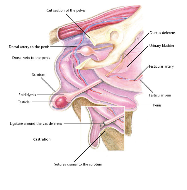
A castration involves the removal of the testicles from within the scrotal sac.
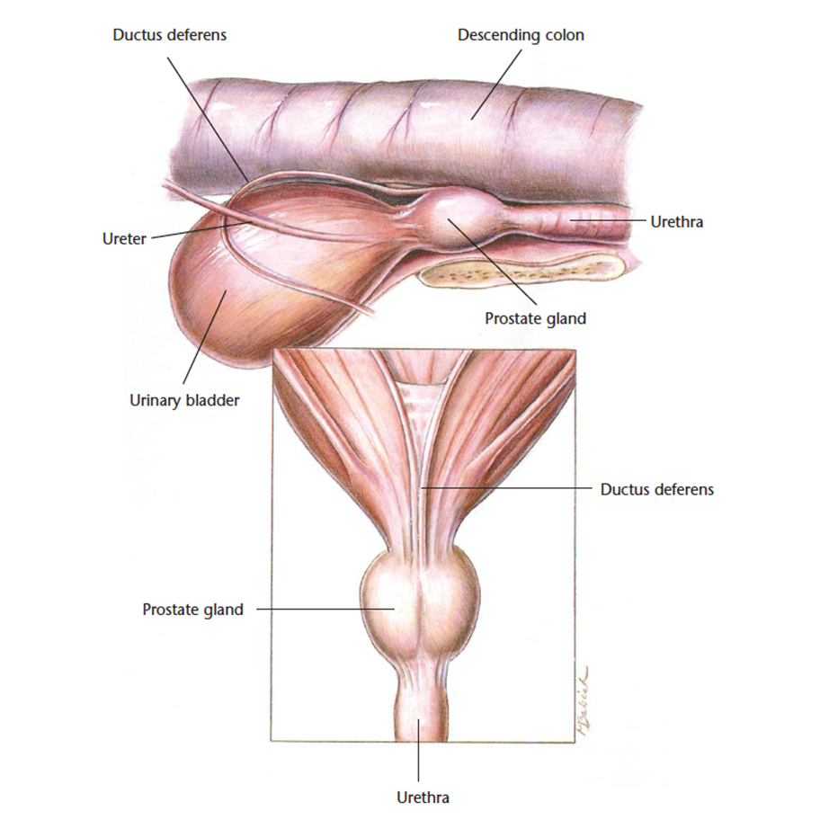
Get access to more anatomical diagrams on the urogenital system.
- Chronic Renal Disease
- Acute Renal Failure
- Bladder Stones
- Urethral Obstruction
- Feline Lower Urinary Tract Disease
- Benign Prostatic Hyperplasia
- Ovariohysterectomy
- Pyometra
- Castration
- Testicular Tumors
Simply register online and once approved, gain access within a few minutes.
JOIN NOWNervous system
The nervous system is responsible for the transmission of messages to and from the brain and spinal cord. The spinal column is protected by the boney spinal vertebrae.
The nervous system includes the:
- Brain
- Spinal cord
- Nerves
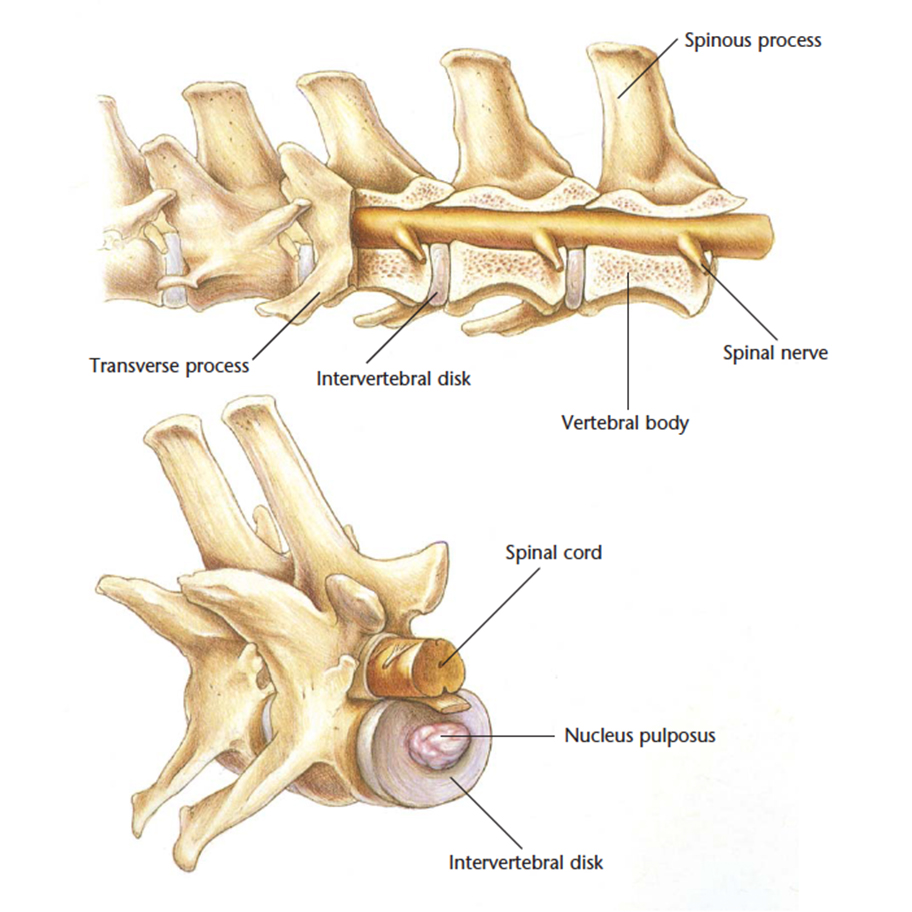
NEUROLOGICAL HANDOUTS
Do you ever see cases of Intervertebral disc disease or cognitive dysfunction? Help your customers understand these complex conditions with the VetCheck handouts.
Simply register online and gain access within a few minutes.
LEARN MOREEye
The eye is responsible for collecting light from the environment and converting this into an image in a three dimensional, moving image.
The eye is made up of the:
- Cornea
- Iris
- Ciliary Body
- Vitreous Body
- Retina
- Lens
- Anterior Chamber
- Optic disk
- Optic Nerve
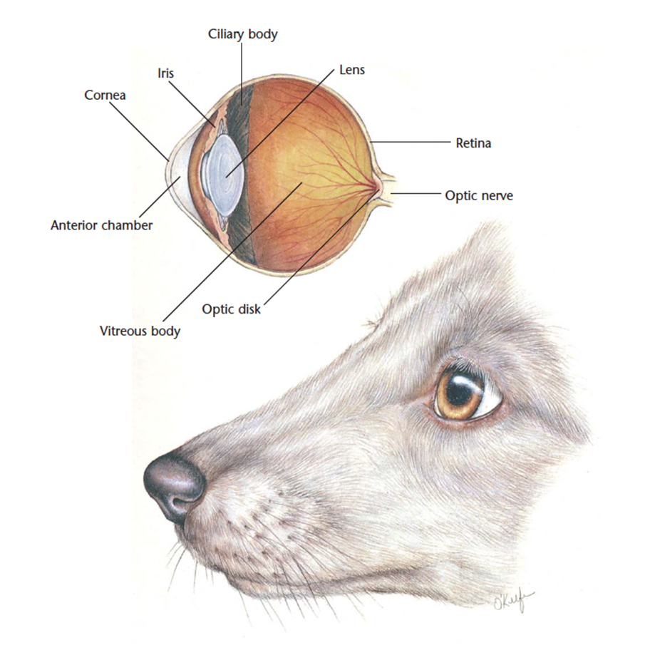
Common eye condition terminology
Here are some common eye condition terms and their meanings:
| Conjunctivitis | Inflammation of the pink tissue inside the eyelids. |
| Uveitis | Inflammation of the middle layer of the eyeball. Represented by eye redness, pain and poor vision. |
| Corneal ulcer | Painful hole in the cornea (the clear membrane on the front of the eye). |
| Keratitis | Inflammation of the cornea. |
| Glaucoma | Increased pressure within the eyeball that can lead to sudden blindness. This is an emergency situation. |
| Lens luxation | Movement of the eye lens out of normal position. |
| Cherry eye | Permanent exposure of the third eyelid. |
| Dry eye | Chronic lack of sufficient eye lubrication that results in irritation of the eye. |
| Retinal detachment | Where the retina comes away from the back wall of the eye. This is an emergency situation. |
| Entropion | The rolling in of the eyelids where the eyelashes constantly rub on the cornea, causing irritation and ulceration. |
| Distichia | Eyelashes that grow from an unusual spot and causes irritation and ulceration. |
| Retinal Dysplasia | Abnormal retinal development that can lead to retinal detachment. |
Breed predispositions
Some dog breeds are prone to eye conditions. Knowing if the dog is genetically predisposed can help pick up early signs of a problem and prevent blindness. Annual vet visits with an eye examination will also help pick up early changes and determine whether further eye examinations are required. This is particularly important for dogs that will be put into a breeding program. Here are some common eye conditions and some breed predispositions*:
| Progressive Retinal Atrophy | Australian Cattle Dog, Collie, Dachshund, Irish Setter, Irish Wolfhound, Lhasa Apso, Labrador Retriever, Miniature Schnauzer, Poodle, Golder Retriever, Cocker Spaniel, Springer Spaniel, Tibetan Spaniel, Welsh Corgi |
| Glaucoma | Maltese, Chinese Crested, Basset Hound, Shiba Inu, Retriever, Siberian Husky, Cocker Spaniel, Springer Spaniel, Spanish Water Dog. |
| Hereditary Cataracts | Bichon Frise, Alaskan Malamute, Australian Shepherd, Boston Terrier, Cavalier King Charles Spaniel, German Shepherd Dog, Giant Schnauzer, Irish Setter, Red Setter, Miniature Schnauzer, Old English Sheepdog, Poodle, Golden Retriever, Labrador Retriever, Siberian Husky, Cocker Spaniel, Springer Spaniel, Staffordshire Bull Terrier |
| Primary Lens luxation | Border Collie, Bull Terrier, Fox Terrier, Jack Russell |
| Collie Eye Anomaly | Australian Shepherd, Border Collie, Shetland Sheepdog |
| Cherry Eye | Bull dog |
| Dry Eye | Cavalier, Chinese Crested |
| Entropion | Chow Chow, Cocker Spaniel, Golden Retriever |
| Retinal Dysplasia | Bedlington Terrier, Cavalier King Charles Spaniel, Golden Retriever, Cocker Spaniel, Springer Spaniel, Labrador Retriever |
* This is not a comprehensive list of genetic eye conditions or breeds.
For a comprehensive list of genetic diseases by dog breed, visit the Orthopaedic Foundation for Animals
Common eye tests that can be performed at the vets:
| Schirmer tear test | Used to meature the tear production. Normal tear production is 10-15mm in one minute. |
| Swab | Sample collection to investigate foreign cells, bacteria or viruses. |
| Fluorescein staining | To check for ulcers that will absorb the stain and fluoresce. It can also be use to check the functioning of the tear duct, where the stain will appear from the nostrils within 5-15minutes if healthy. |
| Tonometry | Tests the pressure within the eye. |
| Gonioscopy | Tests the drainage angle of the eye. |
| Imaging techniques | The use of radiographs, ultrasound or MRI to investigate diseases of the eye or surrounding tissue. |
| DNA swab | The collection of cheek cells or blood to investigate genetic disorders via specific genetic markers. |
Get access to more anatomical diagrams on the eye.
- Normal eye
- Nusclear sclerosis
- Cataracts
- Glaucoma
- Corneal ulceration
Simply register online and once approved, gain access within a few minutes.
JOIN NOWReferences
Garcia-Retamero, R., & Cokely, E.T. (2013). Communicating health risks with visual aids. Current Directions in Psychological Science, 22 (5), 392-399.
These illustrations are available with permission by the copyright owner, Hill's Pet Nutrition, from the Atlas of Veterinary Clinical Anatomy. This illustration should not be downloaded, printed or copied except for non-commercial use.
© Hill's Pet Nutrition Pty Ltd.
Interested in a product tour?
In this demo, we will show you how VetCheck can make your life easier and grow your practice through better client engagement.
Book a 15-minute Demo